
A 12-lead ECG placement guide ensures accurate electrode positioning for optimal heart electrical activity measurement, crucial for precise diagnosis and monitoring of cardiac conditions․
1․1 Overview of 12-Lead ECG
A 12-lead ECG is a comprehensive tool for assessing the heart’s electrical activity, providing a detailed view of cardiac function from multiple angles․ It involves placing 12 electrodes on the body, including limb leads, precordial leads, and augmented limb leads, to capture a wide range of electrical signals․ This method allows healthcare professionals to detect arrhythmias, ischemia, and structural heart abnormalities with high accuracy․ The 12-lead ECG is widely used in clinical settings, emergency rooms, and ambulatory care to diagnose and monitor cardiac conditions․ Its versatility makes it an essential diagnostic tool for both acute and chronic heart diseases․ Proper electrode placement and interpretation are critical for obtaining accurate and reliable results, ensuring effective patient care and management․ This section provides a foundational understanding of the 12-lead ECG system and its significance in cardiovascular assessment․
1․2 Importance of Accurate Placement
Accurate electrode placement is critical for obtaining reliable 12-lead ECG results, as improper positioning can lead to misdiagnosis or delayed treatment․ Even slight deviations from standard positions can cause significant changes in waveform interpretation․ For instance, misplaced limb leads may alter axis determination, while incorrect chest electrode placement can obscure signs of myocardial ischemia․ Ensuring correct placement minimizes artifacts and electrical interference, providing a clear representation of the heart’s electrical activity․ This accuracy is vital for timely and effective patient care, especially in emergency situations where rapid diagnosis is crucial․ Proper training and adherence to established guidelines are essential to maintain consistency and reliability in ECG recordings, ensuring optimal patient outcomes and informed clinical decision-making․

Preparation for 12-Lead ECG
Preparation involves proper patient positioning, skin cleaning, and electrode application․ Ensure the skin is dry and free from oils or lotions for optimal signal quality․
2․1 Patient Preparation
Patient preparation is crucial for accurate 12-lead ECG results․ Ensure the patient is in a comfortable, supine position with arms relaxed at their sides and legs uncrossed․ Remove any jewelry or clothing that may interfere with electrode placement․ Clean and dry the skin thoroughly to ensure proper electrode adhesion and signal quality․ Shave hairy areas if necessary to avoid interference․ Keep the room calm and minimize movement to reduce artifacts․ Ensure the patient remains still and avoids talking during the procedure․ Proper preparation helps in obtaining clear and reliable ECG readings, essential for accurate diagnosis and monitoring of cardiac conditions; Always follow standardized protocols to maintain consistency and patient safety․
2․2 Skin Preparation and Electrode Types
Proper skin preparation is essential for obtaining high-quality 12-lead ECG readings․ Clean and dry the skin thoroughly to remove oils, lotions, or perspiration, as these can interfere with electrode adhesion and signal quality․ Shave hairy areas if necessary to ensure proper contact․ Use a mild abrasive or alcohol wipe to gently abrade the skin, enhancing electrode conductivity․ Select appropriate electrodes, such as silver/silver-chloride or disposable gel electrodes, which provide optimal signal capture․ Ensure electrodes are securely attached to avoid movement artifacts․ For challenging skin conditions, alternative electrodes or adhesive enhancers may be used․ Proper skin preparation and electrode selection are critical for accurate ECG interpretation and reliable diagnostic outcomes․ Always follow manufacturer guidelines for electrode application to ensure consistency and patient safety․
Step-by-Step Placement Guide
A step-by-step guide for 12-lead ECG placement ensures proper electrode positioning․ Follow precise anatomical landmarks for limb, chest, and augmented leads to achieve accurate and consistent results․
3․1 Limb Lead Placement (Arms and Legs)
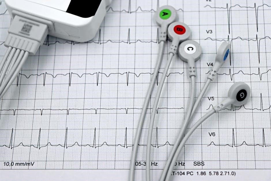
Limb lead placement involves attaching electrodes to the arms and legs to capture the heart’s electrical activity from different angles․ The right arm (RA) and left arm (LA) electrodes are placed mid-way on the inner aspects of the arms, just above the wrists․ Similarly, the right leg (RL) and left leg (LL) electrodes are positioned mid-way on the inner thighs, just above the ankles․ Proper positioning ensures accurate readings of the heart’s electrical axis and helps in identifying potential limb lead reversals․ Chest leads (V1-V6) are then placed across the precordium to complete the 12-lead setup․ Accurate placement is critical to avoid artifacts and ensure reliable ECG interpretations․ Always follow anatomical landmarks for consistency and optimal signal quality․
3․2 Precordial Lead Placement (Chest)
Precordial leads (V1-V6) are placed directly on the chest to capture the heart’s electrical activity from a horizontal plane․ V1 and V2 are positioned at the 4th intercostal space, near the right and left sternal borders, respectively․ V3 is placed midway between V2 and V4․ V4 is located at the 5th intercostal space in the mid-clavicular line․ V5 and V6 are placed horizontally to V4, with V5 in the anterior axillary line and V6 in the mid-axillary line․ Accurate placement is critical to ensure proper waveforms and avoid artifacts․ Use anatomical landmarks to guide positioning, and ensure good skin contact for clear signals․ Proper alignment of these leads helps in identifying conditions like myocardial infarction or bundle branch blocks․ Chest leads complement limb leads to provide a comprehensive view of the heart’s electrical activity․
3․3 Augmented Limb Leads
Augmented limb leads (aVR, aVL, and aVF) are derived from the standard limb electrodes and provide additional views of the heart’s electrical activity․ aVR is placed on the right arm, aVL on the left arm, and aVF on the left leg․ These leads are crucial for detecting lateral and inferior wall abnormalities․ aVR is oriented towards the right shoulder, aVL towards the left shoulder, and aVF towards the lower extremities․ Proper placement ensures accurate waveforms, aiding in the diagnosis of conditions like bundle branch blocks or ventricular hypertrophy․ Augmented leads enhance the diagnostic capability of the 12-lead ECG by offering complementary views to the standard limb leads․ Their correct positioning is essential for comprehensive cardiac assessment and accurate interpretation of ECG tracings․
3․4 Additional Leads and Modifications
Additional leads and modifications are sometimes used to enhance the diagnostic value of a 12-lead ECG․ These include right-sided precordial leads (V1R, V2R, etc․) for detecting right ventricular hypertrophy or infarction․ Posterior leads (V7, V8, V9) may be added to assess the posterior wall of the left ventricle․ Modified leads like the Lewis lead (S5 electrode placement) are used to detect atrial fibrillation more effectively․ These supplementary leads provide more comprehensive coverage of the heart’s electrical activity․ Proper placement and documentation of these leads are essential for accurate interpretation․ They are particularly useful in specific clinical scenarios where standard leads may not provide sufficient information․ Always follow established guidelines to ensure consistency and reliability in ECG interpretation when using additional or modified leads․

Troubleshooting Common Issues
Common issues in 12-lead ECG include electrode artifacts, electrical interference, and improper placement․ Identifying and correcting these problems is essential for accurate ECG interpretation and patient care․
4․1 Identifying and Correcting Artifacts
Artifacts in 12-lead ECG traces are unwanted signals that distort readings, often due to poor electrode placement, skin preparation issues, or external interference․ Identifying artifacts involves recognizing unusual patterns or noise in the tracing․ To correct them, ensure electrodes are properly positioned and secured, with good skin contact․ Replace faulty electrodes and use appropriate types for patient conditions․ Minimize electrical interference by shielding equipment and avoiding nearby devices․ Regularly inspect leads and cables for damage; Proper correction ensures accurate ECG interpretation, critical for diagnosing cardiac conditions effectively․ Consistency in addressing artifacts improves the reliability of ECG results, aiding in timely and accurate patient care;
4․2 Managing Electrical Interference
Electrical interference is a common issue that can disrupt 12-lead ECG readings, leading to inaccurate results․ Sources include nearby electronic devices, fluorescent lighting, and poor equipment grounding․ To manage interference, ensure all ECG equipment is properly grounded and use shielded cables to reduce external noise․ Position the ECG machine away from potential sources of interference, such as monitors or other medical devices․ Additionally, verify that electrodes are correctly placed and secured to minimize signal disruption․ Regularly inspect and maintain equipment to ensure optimal performance․ By addressing these factors, healthcare professionals can reduce electrical interference and obtain clear, reliable ECG tracings essential for accurate cardiac assessments and diagnoses․

Interpretation of 12-Lead ECG
The 12-lead ECG interpretation is vital for diagnosing heart conditions, focusing on P waves, QRS complexes, and T waves to assess rhythm, axis, and signs of ischemia or infarction accurately․
5․1 Basic Interpretation Techniques
Mastering basic 12-lead ECG interpretation begins with analyzing the rate, rhythm, axis, and waveform components․ Start by measuring the heart rate and identifying the rhythm’s regularity or irregularity․ Assess the P wave, QRS complex, and T wave to evaluate atrial and ventricular depolarization․ Determine the electrical axis to detect deviations, which may indicate conditions like left or right ventricular hypertrophy․ Examine the ST segment and T wave for signs of ischemia, injury, or infarction․ Measure intervals, such as the PR, QRS, and QT intervals, to identify conduction abnormalities․ Recognize patterns like bundle branch blocks or hypertrophy․ Interpret findings in the clinical context to diagnose conditions such as myocardial infarction or arrhythmias․ Accurate interpretation requires understanding normal and abnormal patterns, emphasizing the importance of proper electrode placement and skin preparation for clear tracings․ Additional leads, such as V4R, may be needed for further assessment in specific cases․
5․2 Common Pitfalls in Interpretation
Common pitfalls in 12-lead ECG interpretation include misreading artifacts as pathological findings and overlooking subtle changes․ Improper electrode placement can cause misleading ST segment deviations or axis shifts, mimicking conditions like ischemia or hypertrophy․ Failure to consider the patient’s clinical history may lead to incorrect diagnoses․ Overreliance on computer interpretations without manual verification is another pitfall, as algorithms may misclassify rhythms․ Additionally, ignoring baseline wander or electrical interference can obscure key details․ Mismeasurement of intervals, such as the QT interval, can result in false identification of long QT syndrome․ Rookies often confuse benign early repolarization with acute coronary syndromes․ It’s crucial to avoid these errors by ensuring accurate electrode placement, minimizing artifacts, and correlating ECG findings with patient symptoms and other diagnostic tools․ Regular practice and continuous education help improve interpretation accuracy and reduce these common pitfalls․ Staying vigilant is key to avoiding these missteps and ensuring reliable ECG assessments․

Clinical Applications
The 12-lead ECG is crucial for diagnosing myocardial infarction, assessing arrhythmias, and monitoring cardiac conditions, providing comprehensive insights into heart health and aiding in timely interventions․
6․1 Diagnosing Myocardial Infarction
The 12-lead ECG is a cornerstone in diagnosing myocardial infarction (MI), enabling early detection of ischemic changes․ Accurate electrode placement ensures precise readings of ST-segment elevations or depressions, T-wave inversions, and Q-waves, which are critical for identifying MI․ Lead placement helps localize the infarct to specific coronary artery territories, such as the anterior, inferior, or lateral walls of the heart․ Timely interpretation of these patterns allows for prompt interventions, including reperfusion therapy․ Proper placement and interpretation are vital for distinguishing between ST-elevation MI (STEMI) and non-ST-elevation MI (NSTEMI), guiding urgent care decisions․ Additionally, the 12-lead ECG aids in monitoring post-MI complications, such as arrhythmias or bundle branch blocks, ensuring comprehensive patient management․
6․2 Assessing Arrhythmias
The 12-lead ECG is essential for accurately detecting and assessing cardiac arrhythmias, providing a comprehensive view of the heart’s electrical activity․ Accurate electrode placement ensures clear waveform detection, crucial for identifying irregular heart rhythms such as atrial fibrillation, ventricular tachycardia, or supraventricular tachycardia․ By analyzing P-wave morphology, PR intervals, and QRS complexes, clinicians can determine the origin and type of arrhythmia․ The 12-lead ECG also helps differentiate between benign and life-threatening rhythms, guiding immediate treatment decisions․ Proper placement minimizes artifacts, ensuring precise rhythm interpretation․ This tool is vital for monitoring arrhythmias in acute and chronic settings, aiding in the management of conditions like atrial flutter or bundle branch blocks․ Clear ECG waveforms enable timely interventions, improving patient outcomes in arrhythmia care․
6․3 Monitoring Cardiac Conditions
The 12-lead ECG is a critical tool for monitoring cardiac conditions, enabling healthcare providers to track the progression of heart disease and assess treatment efficacy․ Accurate electrode placement ensures consistent and reliable readings, crucial for detecting subtle changes in cardiac function over time․ This method is particularly valuable for monitoring conditions such as coronary artery disease, heart failure, and cardiomyopathies․ By analyzing serial ECGs, clinicians can identify patterns of ischemia, injury, or infarction, guiding timely interventions․ The 12-lead ECG also aids in evaluating the effectiveness of therapies, such as revascularization or medication adjustments․ Regular monitoring with this tool enhances patient care by providing actionable insights into cardiac health, supporting long-term management and improving outcomes for individuals with chronic heart conditions․

Best Practices
Adhering to best practices in 12-lead ECG placement ensures consistency, accuracy, and patient safety․ Proper electrode placement, skin preparation, and equipment calibration are essential for reliable results and effective monitoring of cardiac conditions․
7․1 Ensuring Consistency
Consistency in 12-lead ECG placement is critical for accurate and reproducible results․ Standardized electrode positions and preparation techniques minimize variability, ensuring reliable data across different patients and sessions․ Using anatomical landmarks as reference points helps maintain uniformity․ Regular training and adherence to established protocols are essential for healthcare professionals․ Calibrating equipment and following manufacturer guidelines further enhance consistency․ By maintaining these standards, healthcare providers can ensure that ECG readings are precise and comparable, aiding in accurate diagnoses and effective patient care․ Consistency also supports continuity in monitoring cardiac conditions over time, enabling better tracking of changes and responses to treatment․ Standardization is key to optimizing the diagnostic value of 12-lead ECGs․
7․2 Documentation and Reporting
Accurate documentation and reporting are vital for effective 12-lead ECG interpretation and patient care․ Clear records of electrode placement, patient demographics, and ECG results ensure continuity and accountability․ Standardized reporting formats help healthcare providers quickly identify key findings․ Documentation should include details like date, time, and any relevant clinical context․ Proper reporting involves highlighting abnormal findings and correlating them with patient symptoms․ Clarity and precision in documentation prevent misinterpretation and support informed decision-making․ Regular audits of ECG reports can improve quality and consistency․ Thorough documentation also serves as a legal and clinical reference, ensuring transparency in patient care․ By maintaining accurate and detailed records, healthcare professionals can provide high-quality, patient-centered care․ Effective documentation and reporting are essential for optimizing ECG utility in diagnosis and monitoring․

Resources for Further Learning
For deeper understanding, explore recommended books, online courses, and tutorials on 12-lead ECG placement․ Illustrated guides and practice quizzes enhance learning and proficiency․
Enhance your knowledge with books like “ECG Fundamentals” and “Mastering 12-Lead ECG Placement․” These resources offer detailed guides, illustrations, and practice questions to improve your skills․ They are essential for both beginners and experienced professionals seeking to refine their understanding of accurate electrode placement and interpretation techniques․ Online platforms like Coursera and Udemy offer comprehensive courses on 12-lead ECG placement․ These courses provide interactive modules, video demonstrations, and quizzes to enhance learning․ They cover topics such as electrode placement, skin preparation, and troubleshooting common issues․ Many tutorials include real-world case studies, allowing learners to practice interpreting ECG readings․ Additionally, websites like Quizlet offer flashcards and study guides for memorizing key terms and concepts․ These resources are ideal for healthcare professionals and students seeking flexible, self-paced learning opportunities․ By leveraging these tools, individuals can master 12-lead ECG placement and improve their diagnostic skills effectively․ This guide emphasizes the importance of accurate 12-lead ECG placement for reliable cardiac assessments․ Proper electrode positioning ensures precise measurements of the heart’s electrical activity․ Key points include meticulous skin preparation, correct anatomical placement of limb and precordial leads, and minimizing artifacts․ Understanding augmented limb leads and troubleshooting common issues enhances diagnostic accuracy․ Consistency in electrode placement and adherence to best practices are crucial for optimal ECG interpretation․ Regular practice and reference to detailed guides or courses can improve proficiency․ By following these principles, healthcare professionals can ensure high-quality ECG readings, enabling timely and accurate diagnoses of various cardiac conditions․ Continuous learning and adherence to standardized protocols are essential for mastering 12-lead ECG placement․ Effective 12-lead ECG placement is foundational for accurate cardiac assessments․ Mastery requires attention to detail, proper skin preparation, and precise electrode positioning․ Consistency across placements ensures reliable data, critical for diagnosing conditions like myocardial infarction or arrhythmias․ Minimizing artifacts and electrical interference enhances recording quality․ Regular practice and adherence to best practices are vital for proficiency․ Utilizing resources like guides or courses can further refine skills․ By prioritizing accurate placement, healthcare professionals contribute to better patient outcomes through timely and precise diagnoses․ Continuous learning and adherence to standardized protocols ensure optimal ECG readings, making 12-lead ECG a cornerstone of modern cardiac care․ Effective placement is not just a technical skill but a commitment to patient-centered care․8․1 Recommended Reading Materials

8․2 Online Courses and Tutorials
9․1 Summary of Key Points
9․2 Final Thoughts on Effective Placement